Physique et Imagerie Optique
Club PIO de la SFO
participe au 11ème congrès national OPTIQUE BFC 2026.
Du 6 au 10 juillet 2026 à Dijon.

Présentation du Club
Le domaine de l'imagerie optique a connu ces dernières années des progrès conceptuels et technologiques importants (superrésolution, imagerie 3D, imagerie plénoptique, holographie numérique, compressive sensing,...), en rupture avec les approches « conventionnelles » de conception des systèmes ou de traitement de l'information. Ces progrès ont été rendus possibles par la miniaturisation et la démocratisation des capacités de calcul, mais surtout grâce à l’interaction toujours plus grande entre la physique de l'image, la conception optique, le traitement du signal et des images, et le développement de nouvelles technologies de capteurs.
Le domaine de l’imagerie optique se situant donc plus que jamais au carrefour de ces disciplines, le Club Physique et Imagerie Optique se donne pour mission de favoriser les interactions scientifiques entre les différents acteurs académiques et industriels du domaine de l’imagerie optique non conventionnelle, et de rapprocher ces communautés scientifiques naturellement connexes (Physique de l'image et traitement de l’information par exemple).
Dans ce cadre, les « Journées Imagerie Optique Non-Conventionnelle » (JIONC) organisées annuellement au printemps visent à réunir les chercheurs et ingénieurs de ces différents domaines, afin d'échanger sur les plus récents développements de systèmes ou de traitements pour l'imagerie « non-conventionnelle » et d’évaluer leurs applications potentielles.
Presentation of COLOQ in English
These last years, the domain of optical imaging has experienced tremendous technological and conceptual breakthroughs (superresolution, 3D imaging, light-field imaging, digital holography, compressive sensing,…), with “conventional” approaches of system design or information processing.
Such progress was made possible by the miniaturization and democratization of computing resources, but mostly by the interaction between the physics of image, optical design, signal/image processing, and the development of new sensor technologies.
The domain of optical imaging being more than ever at the crossroad of these fields, the Physics and Optical Imaging Club aims at stimulating the scientific interaction between academic and industrial players of the non-conventional optical imaging domain.
In that context, the annual Non-Conventional Optical Imaging workshop (JIONC) aims at gathering researchers and engineers of these fields, to exchange about most recent developments of systems or processing strategies for “non-conventional” imaging, and evaluate their potential applications.
Charter of the PIO Club of the SFO
Thématiques:
- Modalités d'imagerie non conventionnelles : imagerie hyperspectrale, polarimétrique, holographique, interférométrique, multimodale, thermique, teraHertz, optique de l’extrême (X-UV)…
- Conception d'imageurs innovants : compressive sensing, imagerie 3D/plénoptique, haute résolution, co-conception, …
- Méthodes de traitement en imagerie non conventionnelle : débruitage, déconvolution, segmentation, détection/classification, séparation de sources, ...
- Approches « problèmes inverses » pour l’imagerie
- Imagerie biomédicale : microscopie, OCT, imagerie multi-photonique, super-résolution, imagerie dans les milieux diffusants, …
- Applications de ces systèmes d’imagerie : télédétection, contrôle industriel, défense, astronomie, sciences du vivant, …
Topics:
- Unconventional imaging modalities: hyperspectral, polarimetric, holographic, interferometric, multimodal, thermal, teraHertz, X-UV
- Novel imaging systems design: compressive sensing, 3D/plenoptic imaging, high resolution, co-design,...
- Unconventional image processing: denoising, deconvolution, segmentation, detection/classification, source separation,...
- Inverse problems in imaging
- Biomedical imaging: microscopy, OCT, non-linear imaging, super-resolution, imaging in turbid media,...
- Application of imaging systems: teledetection, industrial inspection, defence, astronomy, life science, ...
Comité Scientifique du club PIO
Président :
Julien FADE (Institut Fresnel)
- Emmanuel Bossy, LiPhy, Université Grenoble-Alpes
- Xavier Briottet, ONERA-DOTA, Toulouse
- Julien Fade, Institut Fresnel, École Centrale Méditerranée
- Alexandra Fragola, ESPCI, Paris Sorbonne Université
- Luc Leviandier, Thales R&T, Palaiseau
- Tatiana Novikova, LPICM, École Polytechnique, Palaiseau
- Jérôme Vaillant, CEA Leti, Université Grenoble-Alpes
- Nicolas Verrier, IRIMAS, Univ. Haute-Alsace, Mulhouse
Légende/Caption
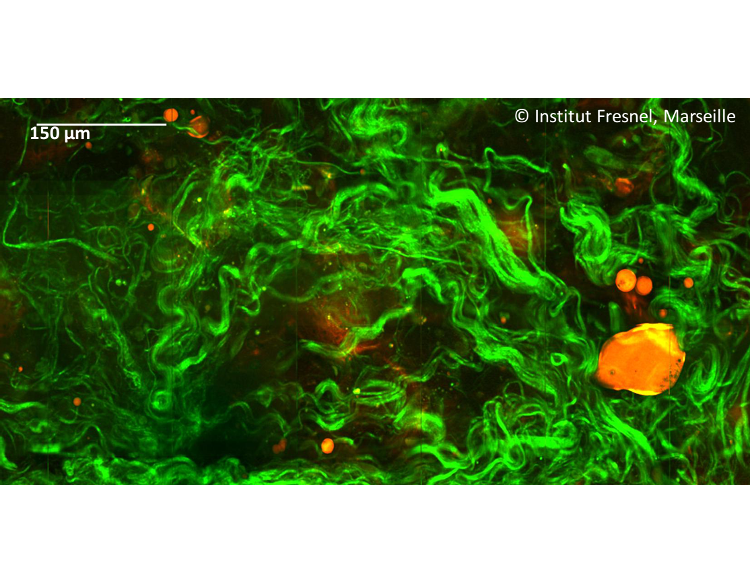
(1) Image non-linéaire dans des tissus biologiques (colon). En vert: collagène, par génération de second harmonique (SHG). En rouge: imagerie vibrationnelle par diffusion cohérente Raman anti-Stokes (CARS) des liaisons CH2 dans les amas lipidiques.
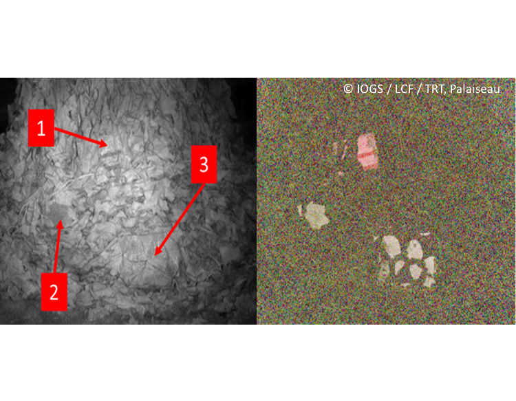
(2) Imagerie polarimétrique pour la détection de cibles à longue portée. Objets métalliques dissimulés par un filet de camouflage. (a) Image active infrarouge classique. (b) Image de contraste polarimétrique d’états orthogonaux.
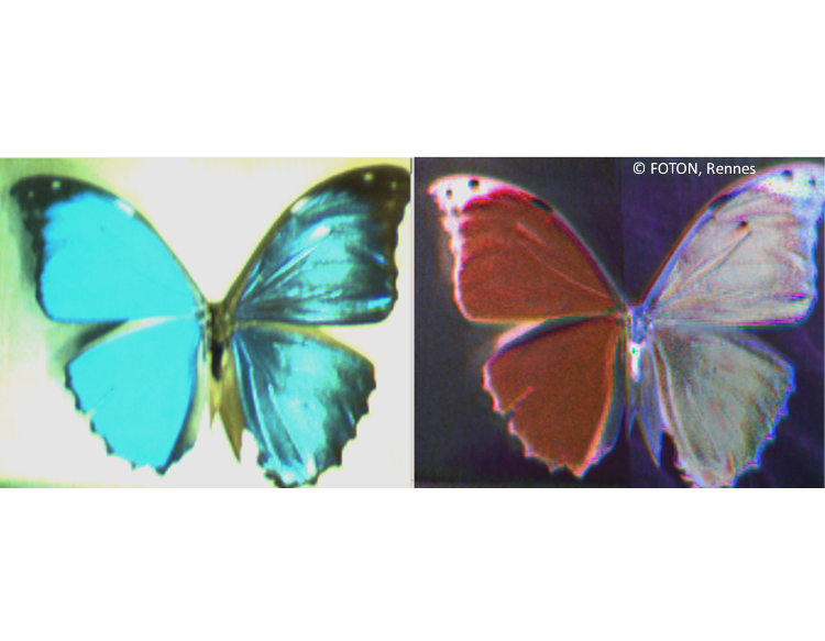
(3) Imagerie spectro-polarimétrique d’un spécimen de papillon morpho (aile droite) et de sa reproduction photographique (aile gauche). À gauche : image d’intensité RGB standard ; à droite : image de contraste polarimétrique d’états orthogonaux RGB.
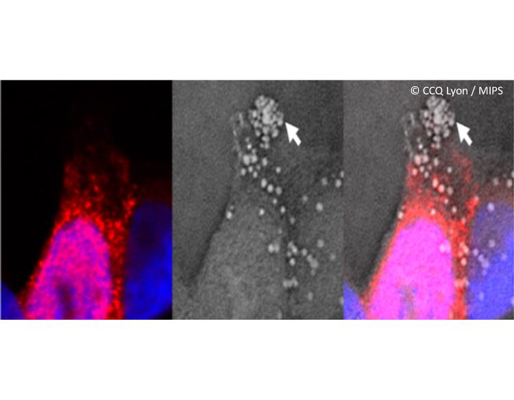
(4) Image multimodale de cellule humaine A549 infectée par le virus H3N2. À gauche : microscopie de fluorescence ; au milieu : microscopie tomographique diffractive ; à droite : fusion des deux canaux d’imagerie.
Crédits : (1) B. Sarri, R. Canonge, H. Rigneault (Institut Fresnel, Équipe MOSAIC) ; (2) N. Verrier, M. Boffety, F. Goudail (LCFIO – IOGS), L. Leviandier, P. Feyneroux (Thales RT); (3) J. Fade, M. Alouini (DOP-UR 1-Rennes) ; (4) A. Béghin, Y. Tourneurs (CCQ Lyon), B. Simon, M. Debailleul (MIPS).
(1) Non-linear image in biological tissues (colon). Green: collagen by second harmonic generation (SHG). Red: vibrational imaging by coherent Raman anti-Stokes diffusion (CARS) of CH2 bonds in lipid aggregates.
(2) Polarimetric imaging for long distance target detection. Metallic objects hidden by a camouflage net. (a) Standard active infrared image. (b) Orthogonal States Polarimetric Contrast image.
(3) Spectro-polarimetric imaging of a morpho butterfly (right wing) and of its printed photograph (left wing). On the left: standard RGB color intensity image; on the right: RGB Orthogonal States Polarimetric Contrast image.
(4) Multimodal imaging of a human A549 cell infected by influenza virus H3N2. On the left: confocal fluorescence microscopy image; in the middle : tomographic diffractive microscopy ; on the right : fusion of the two imaging channels.
Credits : (1) B. Sarri, R. Canonge, H. Rigneault (Institut Fresnel, Équipe MOSAIC) ; (2) N. Verrier, M. Boffety, F. Goudail (LCFIO – IOGS), L. Leviandier, P. Feyneroux (Thales RT); (3) J. Fade, M. Alouini (DOP-UR 1-Rennes) ; (4) A. Béghin, Y. Tourneurs (CCQ Lyon), B. Simon, M. Debailleul (MIPS).
